BCI Stroke Rehabilitation: Does Timing Affect Recovery?
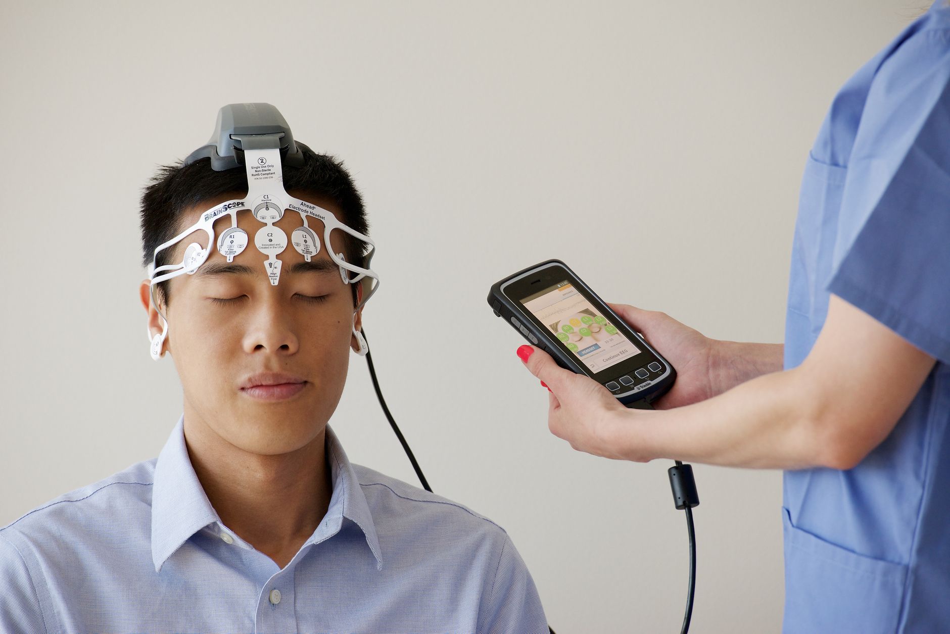
- Varying the timing of motor imagery decoding (1, 2, or 3 seconds) does not improve BCI accuracy.
- BCI accuracy improved in four out of five patients through repeated training.
- Training-induced BCI control did not lead to clinically meaningful motor function improvement.
- Stroke is a network-level disorder, making functional connectivity a key feature for BCI feedback.
Does decoding window timing improve BCI performance?
Changing the onset of the motor‑imagery decoding window does not improve BCI performance or clinical recovery; the study found no significant effect of decoding timing on accuracy, fatigue, or spasticity. This lack of effect was consistent across the varied 1‑, 2‑, and 3‑second windows and did not translate into meaningful motor‑function gains despite participants learning to control the BCI.
Can upper limb rehabilitation benefit from network-based feedback?
Motor‑imagery BCIs are pursued for upper‑limb rehabilitation because many stroke survivors have residual distal impairment that prevents conventional active practice. Current research highlights stroke as a disorder at the network level, making methods that include functional connectivity and EEG signal covariance stronger candidates for network‑based feedback in MI‑BCI post‑stroke. The preprint titled “Single‑Case Design for Network‑based BCI Rehabilitation…” was released by a consortium of French research institutions on 23 October 2026, providing the context for this network‑focused investigation. The researchers examined if patients could tolerate the intensive protocol, if their online BCI accuracy increased during training alongside particular brain activity and network alterations, and if the MI timing exerted a notable overall effect. All participants gave written informed consent in line with the Declaration of Helsinki.
How does motor imagery BCI impact stroke recovery?
Five chronic stroke patients, BCI‑naïve, at a minimum of six months after their first lesion, and exhibiting residual distal upper‑limb deficit (UE‑FMA < 53) with observable active finger flexion (MRC ≥ 2), were recruited for the study. Participants were excluded if they had poor command comprehension (BDAE < 12), fixed flexor spasticity (mAS = 4), a recent botulinum toxin injection, or any contraindication to functional electrical stimulation or EEG recording. Additional eligibility details and the full assessment battery are described in the supplementary methods (Methods S1 and S2.1). All participants underwent 18 BCI training sessions across six weeks, with intervals of one to two days, and subsequently a post‑intervention assessment after three months. Each session started with a short breathing exercise and guided motor imagery, employing a detailed script for the initial three sessions and a shortened version thereafter. Bilateral forearm electromyography was recorded to verify that no muscular contractions occurred during the motor‑imagery phases. The decoding window was a fixed three‑second segment: during rest it covered the first three seconds, while during motor imagery its position depended on the randomized treatment—starting 1 s after cue for a 4‑second MI, 2 s after cue for a 5‑second MI, or 3 s after cue for a 6‑second MI. The timing of the window was randomized according to an alternating‑treatments single‑case experimental design, allowing each patient to serve as their own control. The comprehensive protocol was executed as a functional electrical stimulation‑controlled EEG network‑based MI‑BCI investigation with the same five chronic stroke patients across the 18 uniformly spaced sessions.
What technology powers the BCI system?
The system records EEG data using a 64-channel BrainAmp system with active electrodes sampled at 500 Hz. The setup follows a 10-20 montage referenced to the mastoids, with electrode impedance maintained below 10 kΩ at the start of each session. Researchers applied a 50 Hz notch filter and common average reference, while excluding electrodes FT9, FT10, and Iz from analysis.
The BCI pipeline is implemented in OpenViBE, while MNE-Python handles signal processing and statistical analysis. HappyFeat software handles feature extraction and decoder training, combining spectral power and functional connectivity features to categorize motor imagery states. Functional electrical stimulation is delivered through the FESIA Grasp system on the paretic forearm, calibrated before training to ensure a standardized hand opening.
How did researchers measure patient progress?
Performance was monitored via online BCI accuracy, calculated as the count of correctly classified Rest and motor imagery phases divided by twice the total trial count. To test whether timing affects control, the team used a randomized alternating-treatments design that shifted a fixed 3-second decoding window to 1, 2, or 3 seconds after the cue onset. Offline metrics included run-wise cross-validated accuracy, Rest distinctiveness, and Class stability, with covariance matrices estimated on unreferenced raw EEG.
The system calculated power spectral density over 0.25-second epochs using an autoregressive Burg model of order 24, averaged to create a single measure for the 3-second window. Functional connectivity was measured using imaginary coherence on sliding 0.5-second epochs to suppress zero-lag correlations from volume conduction. Researchers identified the most strongly connected electrodes by calculating node strength, which is the sum of weighted connections incident to each electrode.
Patient state was monitored using a tactile visual scale from 0 to 100, where participants rated mood, stress, and sleep quality before sessions, and fatigue before, during, and after sessions. Spasticity monitoring employed the modified Ashworth scale on the thumb, index, other fingers, and wrist, yielding a global score between 0 and 20. Upper-limb motor function was assessed using the Fugl-Meyer Assessment before the first and last sessions, with all scales repeated at the follow-up visit.
What were the results for the participants?
Four of the five patients showed an increase in online BCI accuracy throughout training. These gains coincided with longitudinal, patient‑specific alterations in local spectral activity and functional connectivity. There was no screening for BCI aptitude applied to the participants.
What are the challenges in BCI rehabilitation protocols?
Standardizing BCI protocols remains difficult because a wide variety of experimental paradigms exist, making it hard to identify the best practices. Researchers often apply fixed parameters to all participants, despite the fact that patients possess substantially different behavioral and neurophysiological characteristics. Factors such as lesion location and extent, the severity of motor deficits, cognitive comorbidities, and the stage of recovery all influence how an individual responds to treatment. These factors also dictate the specific oscillatory frequency band where a patient's response appears and how that response changes throughout a training program. Because event-related desynchronisation trajectories can run in opposite directions within the same group, you cannot rely on group-average effects to describe the progress of an individual patient. While guidance for user training exists for healthy volunteers, no equivalent protocols cover rehabilitation after stroke.
Conventional therapies typically deliver about 32 repetitions per session, whereas animal models suggest that 500 repetitions are necessary to induce cortical synaptic changes. However, you must avoid excessive fatigue and drowsiness, especially since the EEG setup itself consumes 20% of the entire session. Trial duration involves a constant trade-off: longer trials of 3 to 10 seconds or more may better account for motor system alterations, while shorter trials aim to shorten the session and reduce patient fatigue. Including a physical object like a target cup can make the motor imagery task more ecological, while random inter-trial intervals of 1 to 2 seconds help reduce temporal predictability.
The feature‑selection process was repeated at three time points during each session to adapt the classifier to non‑stationarities from mental fatigue, strategy changes, and signal drift. You can adapt to these changes by training the initial classifier on a calibration run, then retraining it after the first two testing runs and again after the fifth run to maintain accuracy.
- Single-Case Design for Network-based BCI Rehabilitation: Individual Learning Trajectories and Effects of Motor Imagery Timing in Stroke Patients — Sorbonne Universite, Paris Brain Institute, ICM, CNRS, Inria, Inserm, AP-HP, Hopital de la Pitie-Salpetriere, Paris, 75013, France, Oct 23, 2026
- Single-Case Design for Network-based BCI Rehabilitation: Individual Learning Trajectories and Effects of Motor Imagery Timing in Stroke Patients — medRxiv, Oct 23, 2026
- Single-Case Design for Network-based BCI Rehabilitation: Individual Learning Trajectories and Effects of Motor Imagery Timing in Stroke Patients — medrxiv.org, Oct 23, 2026
- Walking (Wikipedia — background) — Wikipedia
Add NewsPulse Time as a preferred source on Google
Frequently asked questions
No. According to the study, varying the onset of the motor imagery decoding window (1, 2, or 3 seconds after the cue) had no significant effect on BCI accuracy, patient fatigue, or spasticity. While most patients improved their BCI control over 18 sessions, this did not result in clinically meaningful improvements in their motor function.