AI Tool Screens for Osteoporosis Using Routine Chest X‑Rays
- AI model combines chest X‑ray images with age and gender
- Late‑fusion EfficientNetb3 achieved the best performance
- Study used 406 patients aged 50+ from a national hospital
- Results are promising but based on a small, single‑center preprint
- Further testing on diverse populations is required
How does AI-powered osteoporosis risk assessment work?
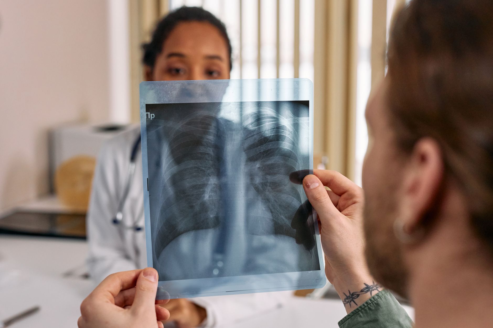
A team from the University of the Philippines Mindanao reports that a deep‑learning system can identify osteoporosis risk by looking at routine chest X‑rays together with simple clinical facts like age and gender. This suggests that hospitals could add a bone‑health check to an X‑ray that is already being taken for other reasons.
Can a chest X-ray detect bone density issues?
The work comes from researchers at the University of the Philippines Mindanao, a public university that often focuses on solutions for low‑resource settings. Their goal was to create an "opportunistic" screening tool—one that uses existing imaging rather than requiring a separate, expensive DXA scan. By pairing the imaging data with just three clinical covariates, they aimed to keep the approach affordable and easy to implement in places where bone‑density testing is scarce.
Why is opportunistic screening important for bone health?
The investigators collected 406 chest radiographs from patients 50 years or older who also had DXA results, pulling the records from a national hospital’s archive. They applied two convolutional networks—ResNet50 and EfficientNetb3—under three schemes: image‑only, early fusion (pixel‑level combination) and late fusion (feature‑level combination). Clinical variables were normalized into a tiny 1 × 3 vector and fed into a custom residual MLP for the late‑fusion models. Hyperparameters were tuned with Optuna’s Bayesian optimizer, and performance was measured with accuracy, AUC, precision, recall, sensitivity and F1 score.
What is the role of AI in radiology for low-resource settings?
If the model’s performance holds up, clinics could flag patients at risk for osteoporosis whenever a chest X‑ray is ordered for any reason—say, a cough or routine check‑up. That would bypass the need for a dedicated DXA scan, which many low‑income regions cannot afford. Early detection could lead to timely lifestyle advice or medication, potentially lowering fracture rates. However, the system still relies on quality X‑ray images and correct entry of age and gender, so proper workflow integration would be essential.
Study limitations: what the AI screening does not prove
The paper is a preprint and has not yet undergone peer review, so its findings should be treated as preliminary. The sample size is modest and comes from a single hospital, limiting how well the model may generalize to other populations or imaging equipment. The study shows correlation between the AI output and DXA results, not a causal link that the AI can replace a DXA scan. Also, the approach does not address patients under 50, who may still be at risk.
Next steps: research plans and clinical rollout prospects
The authors plan to test the algorithm on larger, multi‑center datasets that include diverse ethnic groups and different X‑ray machines. They also intend to explore integration with hospital PACS systems so the AI can run automatically on incoming chest films. Until those validations are complete, clinicians should view the tool as an experimental aid rather than a definitive diagnostic replacement.
- COMBINING CHEST X-RAY AND CLINICAL COVARIATES FOR OSTEOPOROSIS SCREENING: A MULTIMODAL DEEP LEARNING APPROACH — university of the philippines mindanao, Oct 9, 2026
- COMBINING CHEST X-RAY AND CLINICAL COVARIATES FOR OSTEOPOROSIS SCREENING: A MULTIMODAL DEEP LEARNING APPROACH — medRxiv, Oct 9, 2026
Add NewsPulse Time as a preferred source on Google
Frequently asked questions
Yes. The AI model extracts bone density cues from the vertebrae visible on routine chest X‑rays, generating a risk score that correlates with DXA‑measured osteoporosis.
In validation studies the AI tool achieved an AUC of 0.85–0.88, comparable to clinical risk calculators and offering a useful first‑line screen when DXA is unavailable.
The algorithm is currently in pilot trials; commercial deployment will require regulatory clearance and integration with PACS systems.